|
|
Ras Binding Domain
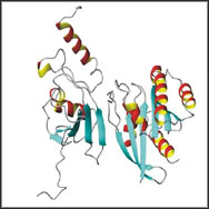
Small GTPase Raf |
| |
|
Cells are able to respond to their extracellular environment by transducing signals received at the me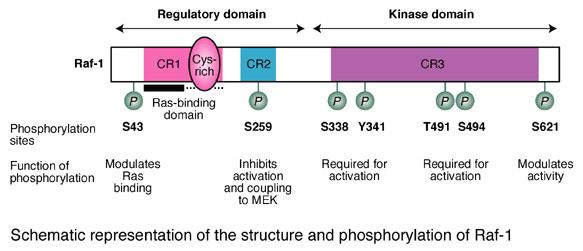 mbrane through cascades of intracellular proteins to physiological effectors. These intracellular signal transduction pathways serve to control key cellular processes including cell proliferation, differentiation, programmed cell death, changes in cell shape and motility, and manifestation of the tumor phenotype. Central to many of these pathways are MAP kinase modules, the archetypal form of which is a cascade of sequentially acting protein kinases named Raf, MEK1/2 and ERK1/2 that are activated downstream of cell surface receptors and the Ras GTPases. Raf-1 is ubiquitously expressed, whereas A-Raf is predominantly found in urogenital tissue and B-Raf shows the highest expression in neural tissue and testis. The three Raf proteins share a common structure consisting of an N-terminal regulatory domain and a C-terminal kinase domain. There are three conserved regions (CR1-3) of homology: in the regulatory domain, CR1 harbours a Ras-binding domain and a cysteine-rich domain, and CR2 is a serine/threonine-rich domain; CR3 is sited in the kinase domain and is required for Raf activity. All three Raf proteins also share common mechanisms of activation and downstream effectors. Activation of Ras by tyrosine kinase autophosphorylation recruits Raf to the membrane where Raf is avtivated and in turn activate the MAPK/ERK pathway by phosphorylating the MAPK/ERK kinase (MEK). mbrane through cascades of intracellular proteins to physiological effectors. These intracellular signal transduction pathways serve to control key cellular processes including cell proliferation, differentiation, programmed cell death, changes in cell shape and motility, and manifestation of the tumor phenotype. Central to many of these pathways are MAP kinase modules, the archetypal form of which is a cascade of sequentially acting protein kinases named Raf, MEK1/2 and ERK1/2 that are activated downstream of cell surface receptors and the Ras GTPases. Raf-1 is ubiquitously expressed, whereas A-Raf is predominantly found in urogenital tissue and B-Raf shows the highest expression in neural tissue and testis. The three Raf proteins share a common structure consisting of an N-terminal regulatory domain and a C-terminal kinase domain. There are three conserved regions (CR1-3) of homology: in the regulatory domain, CR1 harbours a Ras-binding domain and a cysteine-rich domain, and CR2 is a serine/threonine-rich domain; CR3 is sited in the kinase domain and is required for Raf activity. All three Raf proteins also share common mechanisms of activation and downstream effectors. Activation of Ras by tyrosine kinase autophosphorylation recruits Raf to the membrane where Raf is avtivated and in turn activate the MAPK/ERK pathway by phosphorylating the MAPK/ERK kinase (MEK).
It has been confirmed that the minimum structure of the Raf-1 serine/threonine kinase that recognizes active Ras is not Ras-binding domain (RBD, 51-131) but RBD plus the cysteine-rich domain (CRD) (Raf-1[51-220]). In normal NIH 3T3 cells, GFP tagged Raf-1[51-220] showed minimal membrane localization that was enhanced after stimulation with platelet-derived growth factor (PDGF). Mutations within either the RBD (R89L) or CRD (C168S) disrupted the membrane localization of (Raf-1[51-220]GFP), suggesting that both domains contribute to the recruitment of the fusion protein to Ras at the plasma membrane and Raf-1[51-220] is a great tool to visualize activation of Ras-Raf-MEK-ErK pathway
(see the following cartoon for Raf structure). |
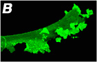
An example
showing the membrane accumulation of RBD of Raf-1 (51-220) after application
of PDGF
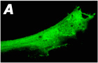  Left
Panel:
RBD+CRD of Raf-1 (51-220) is cloned into a GFP vector. GFP tagged Raf (A) moves to the
cytoplasma after treatment with PDGF (100 ng/ml) (B) while a truncated
Raf (51-131) loses the membrane binding capapctiy (images not
shown). All the images were acquired by Laica SP2 confocal microscopy
and courteously provided by Dr. Zhou from National Institutes of Health
(NIH) Left
Panel:
RBD+CRD of Raf-1 (51-220) is cloned into a GFP vector. GFP tagged Raf (A) moves to the
cytoplasma after treatment with PDGF (100 ng/ml) (B) while a truncated
Raf (51-131) loses the membrane binding capapctiy (images not
shown). All the images were acquired by Laica SP2 confocal microscopy
and courteously provided by Dr. Zhou from National Institutes of Health
(NIH) |
|
|