We use cookies to enhance your browsing experience. Essential cookies are always enabled. By clicking "Accept" or continuing to use this site, you consent to the use of optional analytics and tracking cookies.
Service Quote Request
Please fill out the form below with your detailed inquiry.
Genetically Encoded Calcium Indicators (GECIs) are fluorescent proteins engineered to report intracellular calcium (Ca²⁺) dynamics in living cells. Because Ca²⁺ influx closely accompanies neuronal firing, synaptic activity, muscle contraction, and many signaling pathways, GECIs provide a powerful way to visualize cellular activity in real time.
🧬 How GECIs Work
Most GECIs are based on a calcium-sensitive fluorescent protein whose brightness changes when Ca²⁺ binds.
Classic GCaMP architecture
Circularly permuted GFP (cpGFP)
Calmodulin (CaM) — Ca²⁺ sensor
M13 peptide — CaM-binding domain
Mechanism
Ca²⁺ binds calmodulin
CaM interacts with M13
Conformational change increases GFP fluorescence
Signal intensity reflects intracellular Ca²⁺ levels
🧠 Why Calcium = Activity
In excitable cells (especially neurons):
Action potentials open voltage-gated Ca²⁺ channels
Synaptic activation triggers Ca²⁺ entry or release from ER
Ca²⁺ acts as a universal second messenger
Thus, GECIs provide an indirect but robust readout of cellular activity.
⭐ Major GECI Families
🟢 GCaMP Series (Most Widely Used)
4
Single-color green indicators
High sensitivity and signal-to-noise
Available in multiple kinetics
Examples
Variant
Key Property
Typical Use
GCaMP6s
High sensitivity, slow
Sparse firing
GCaMP6f
Fast kinetics
Rapid activity
GCaMP7/8
Improved brightness & speed
Modern studies
🔴 Red GECIs (e.g., jRGECO, RCaMP)
4
Longer wavelengths penetrate tissue better
Reduced phototoxicity and scattering
Enable multicolor imaging with GFP-based tools
Advantages
Compatible with optogenetics using blue light
Better for deep-brain imaging
Useful for dual-population recordings
🔬 Delivery Methods
GECIs are genetically encoded, so they must be introduced via gene delivery:
Common approaches
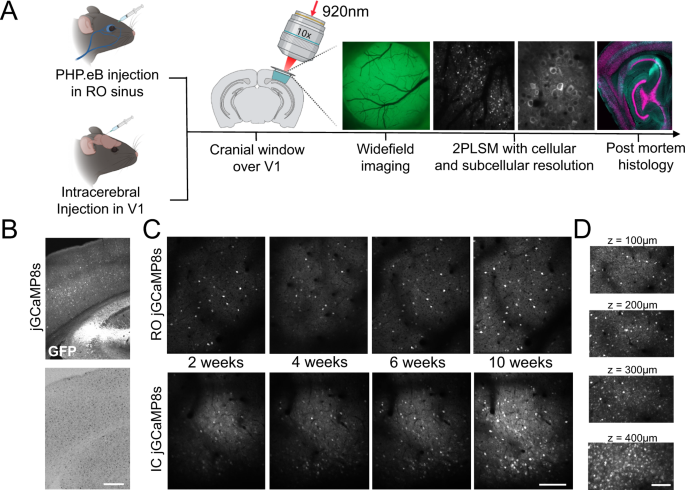
rAAV vectors (most common in vivo)
Lentiviral vectors
Transgenic animals
In utero electroporation
Plasmid transfection (in vitro)
Given your work with viral vectors at SignaGen, rAAV-mediated delivery is typically the gold standard for stable, cell-type-specific expression in animal studies.
🧪 Experimental Modalities
GECIs support multiple recording techniques:
In vivo brain activity
Two-photon microscopy
Widefield imaging
Miniscope (head-mounted) imaging
Fiber photometry
In vitro systems
Cultured neurons
Brain slices
Organoids
Non-neuronal cells
⚠️ Limitations
Indirect measure of electrical activity
Slower than electrophysiology
Signal saturation at high firing rates
Photobleaching and phototoxicity
Calcium buffering can perturb physiology
🧠 Typical Applications
Neural circuit activity mapping
Behavior-linked activity recording
Synaptic plasticity studies
Cardiac and muscle physiology
Immune cell signaling
Drug screening
🏆 Why GECIs Are Transformative
They enable long-term, cell-type-specific, non-destructive recording of activity in living organisms — something impossible with traditional dyes or electrodes alone.